

ПЕРЕДОВЫЕ СТАТЬИ
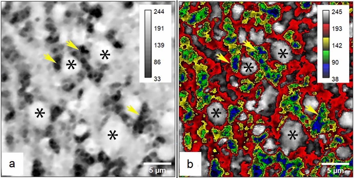
This study examines the methodological aspects of applying machine learning algorithms to explore new opportunities in interpreting inter-neuronal connections. The aim was to demonstrate that the combination of Ilastik and StarDist is effective for the morphometric characterization of giant synaptic terminals in the stratum lucidum of CA3 in the hippocampus of white rats under normal conditions and in the post-ischemic period. Material and methods. Cerebral ischemia in Wistar white rats was modeled by bilateral occlusion of the common carotid arteries (OCCA) for 20 minutes. Animals were studied without intervention (n=6, control) and at 6 hours, 1, 3, 7, 14, and 30 days after OCCA (n=36). Staining with hematoxylin and eosin, Nissl staining with thionine, as well as immunohistochemical reaction for synaptophysin, were used. Numerical density (NDT), sizes, staining intensity, and area of the terminals were determined, and the Ilastik and StarDist plugins were applied on the ImageJ/Fiji platform. Statistical analysis was performed using non-parametric methods in Statistica 8.0. Results. The relative area of the terminals did not differ between the manual method and machine learning. Machine learning provided additional information on numerical density, sizes, and average brightness of the terminals. At 6 hours after OCCA, NDT decreased by 44.3%, but then recovered over 7 days. The average area of the terminals was 16.7% larger at 6 hours and 1 day, but smaller than the control level at 14 days. The brightness of the terminal pixels was inversely proportional to the content of chromogen: it increased at 6 hours and 1 day after OCCA, then returned to control levels. Correlations were observed between the area and brightness of the terminals (R=0.78). Conclusion. The use of the combination of Ilastik and StarDist allowed for accurate assessment of numerical density, sizes, shape, relative area, and staining intensity of synaptic terminals in the hippocampus. Compared to the manual method, the application of machine learning provided significantly more information about the terminals in color immunohistochemical images.
ORIGINAL PAPERS
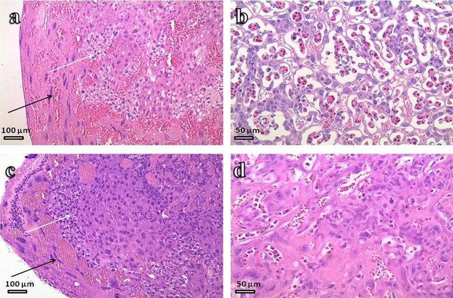
The aim was to characterize morphological and functional changes in the placenta in midpregnancy when modeling experimental preeclampsia and evaluate the model for validity. Material and methods. The study was conducted on 12–14-week-old mice of the inbred CBA, DBA/2, Balb/c lines. Uncomplicated pregnancy was modeled using the ♀CBA×♂Balb/c combination, preeclampsia was reproduced by the ♀CBA×♂DBA/2 combination and the introduction of 0.1 ml of 0.9% NaCl solution of beta-heptylglycoside muramyl dipeptide (C7MDP) intraperitoneally at the dose of 25 μg per 1 animal on days 5 and 7 of gestation (DG). On day 14 of gestation, the frequency of fetal resorption and the level of proteinuria were determined, and the placentas were dissected. Placenta preparations were stained with hematoxylin and eosin. Freshly isolated placenta was also examined using scanning electron microscopy, placenta homogenate was used to measure ex vivo cytokine production using flow cytofluorimetry. After birth of the offspring, the fetuses were weighed on the 1st day of life and the crown-rump length was measured. The obtained results were statistically processed using the Sigma Stat 3.5 program. Results. The proteinuria level was 0.1 (0.0; 0.1) g/l in cases of uncomplicated pregnancy and 1.0 (1.0; 3.0) g/l in pregnancy complicated by preeclampsia. On the 14th day of pregnancy, the resorption rate in cases of the uncomplicated pregnancy group was 12.9%, in group of experimental preeclampsia we revealed 39.34% ones. Morphometric study showed intrauterine growth retardation of the fetuses in the experimental preeclampsia group. Our study of cytokines estimating in placental homogenates showed that pregnancy complicated by experimental preeclampsia was accompanied by significant increase in the level of the proinflammatory cytokine IL-1, as well as decrease in the pleiotropic cytokines IL-4 and IL-6 and anti-inflammatory IL-10. Using light and scanning electron microscopy we found morphological and functional changes in the placenta in experimental preeclampsia reflecting the signs of placental insufficiency. Conclusion. The changes we observed allow us to classify the considered model of preeclampsia as one of the most adequate at present. Having high reproducibility and validity, it can contribute to the search for new diagnostic and therapeutic targets for this pregnancy complication.
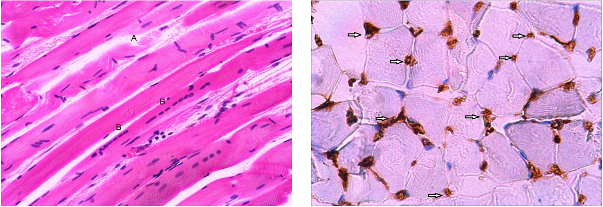
The aim was to study the structural and functional reorganization of the sternocleidomastoid muscle, diaphragm and external intercostal muscles in chronic heart failure (CHF). Material and methods. Three groups of inspiratory muscles were studied: sternocleidomastoid muscle, diaphragm, external intercostal muscles, as well as psoas minor muscle as a control in 24 dissected male cadavers. Based on the presence of CHF and its stage, patients were divided into three groups: without signs of CHF (n=8), with CHF stage I-IIA (n=8), with CHF stage IIB-III (n=8). Histological sections were stained with histological and immunohistochemical techniques (CD34 biomarker expression was detected to determine the activity of angiogenesis and capillary density). Neutral polysaccharides (glycogen) were determined by the PAS reaction. The morphometric study was used to calculate the relative area of endomysial vessels and the diameter of muscle fibers. Results. In patients without signs of CHF, among all the muscles studied, the most pronounced changes were observed in the diaphragm. The highest indicators of the relative area of blood vessels and capillary density on a conditional unit of area were revealed; thickened myosimplasts prevailed and the most intense PAS reaction was revealed. As heart failure increases, the number of capillaries in the diaphragm decreases, glycogen reserves are depleted and the number of thin myosimplasts increases. In external intercostal muscles, the maximum values of relative vascular area and capillary density were observed in CHF stage I-IIA. In all groups, medium-thick myosiplasts prevailed in this muscle. In sternocleidomastoid muscle, the highest indicators of relative vascular area and capillary density were found in the group with CHF stage I-IIA. In CHF stage IIB-III, these indicators did not decrease compared to the group without CHF. The psoas minor muscle study revealed an increase in the relative vascular area in CHF stage IIB-III compared with the group without CHF, which is probably due to venous congestion. Conclusion. The revealed structural and functional reorganization of inspiratory muscles depends on the degree of their participation in the breathing. Each muscle reacts differently to the increase in heart failure with the appearance of signs of compensation and decompensation, depending on the stage of CHF.
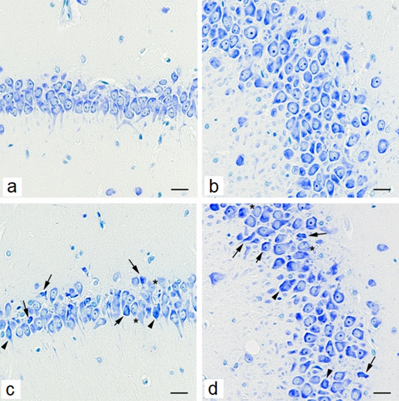
The aim was to study the morphological changes in the visual cortex of the brain in young and old rats treated with a fructose-fat diet (FFD). Material and methods. The study was carried out on male Wistar rats, divided into the following groups: 1st (n=14) – intact 6-month-old rats received a standard diet; 2nd (n=14) – 6-month-old rats received a fructose-fat diet (FFD) for 90 days (from 3 months of age); 3rd (n = 14) – intact 18-month-old rats received a standard diet; 4th (n = 14) – 18-month-old rats received the FFD for 90 days (from 15 months of age). Histological sections were Nissl stained. Immunohistochemical reaction was performed to detect the expression of vascular endothelial growth factor (VEGF). In layers II, IV and V of the primary visual cortex, the percentage of normochromic and altered neurons and the number of gliocytes in 1 mm2 of section were calculated. Differences between groups were determined using the Kruskal–Wallis multiple comparison test. Results. In Morphological changes in the visual cortex in 18-month-old rats were most pronounced in layers IV and V and, in addition to reversible neuronal disorders, were characterized by total chromatolysis and vacuolization of the cytoplasm. In 6-month-old rats on a FFD, the most pronounced increase in hyperchromic neurons with shrinkage was observed in layer IV. In 18-month-old animals, pathological changes in neurocytes were found in all studied layers of the primary visual cortex, and pronounced signs of neuronophagia and gliosis were noted. In 18-month-old intact rats and 6- and 18-month-old rats on a FFD, vascular congestion and perivascular edema and pronounced expression of VEGF were detected. Conclusion. FFD enhances age-related changes in the visual cortex of rats, manifested by vascular disorders, degenerative changes in neurons and glial hyperreactivity.

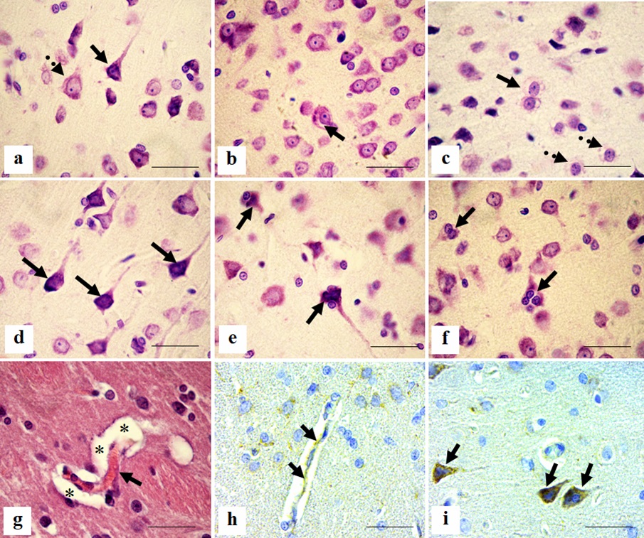
Cholestasis, characterized by stagnation of bile in the liver and impaired excretion of bile into the intestine, causes digestive disorders and the entry of toxic components of bile into the blood and brain. The study of the effect of cholestasis on cholinergic neurons of the cingulate cortex in rats allows us to understand the mechanisms of pathological changes in the brain. The aim was to investigate the effect of cholestasis on cholinergic neurons of cingulate cortex in rats. Material and methods. The study involved 72 outbred white male rats weighing 225±25 g. Subhepatic cholestasis was modeled using the method of L.S. Kizyukevich. The animals of the control group underwent a sham operation. Rats of the control and experimental groups were decapitated on the 2nd, 5th, 10th, 20th, 45th and 90th days. fragments of the cerebral hemispheres were embedded in paraffin and 5 µm thick frontal sections were prepared, stained with antibodies to the choline acetyltransferase (ChAT) protein. Neurons of the cingulate cortex of the rat brain were studied. The number of cholinergic neurons in the preparations was counted, and the relative content of ChAT in their cytoplasm was cytophotometrically estimated. The data were subjected to statistical processing. Results. It was found that, under normal conditions, the number of cholinergic neurons in the parvocellular layer of the cingulate cortex is greater than in the magnopyramidal layer. The content of ChAT in the cytoplasm of the perikarya of neurons in the parvocellular layer decreases 5 and 10 days after cholestasis. With continued cholestasis (on the 10th and 20th days after surgery), the number of cholinergic neurons in the parvocellular layer of the cingulate cortex decreases, indicating their death. The content of ChAT in the cytoplasm of neurons of the magnopyramidal layer decreases only by the 10th day of cholestasis. In this case, the maximum loss of cholinergic neurons is detected on the 45th day. Cholinergic neurons of the parvocellular layer respond faster to cholestasis and the surviving neurons normalize earlier after its disappearance, compared with neurons of the magnopyramidal layer of the cingulate cortex. Conclusion. A decrease in the content of ChAT, indicating a violation of acetylcholine synthesis, and the loss of cholinergic neurons in the cingulate cortex may underlie the cognitive deficit observed in patients with cholestasis.

The aim was to establish the nature of liver remodeling in hyperglycemia and insulin resistance in leptin-resistant db/db mice and to study the possibility of reversing the identified structural changes during glucose metabolism correction using systematic administration of recombinant apolipoprotein A-I (rApoA-I). Material and methods. The structural organization of the liver was studied in db/db mice (n=46) at the age of 10, 12 and 24 weeks, including weekly subcutaneous administration of rApoA-I (at a dose of 7 mg/kg body weight) as a glucose metabolism corrector from the age of 8 weeks. C57/Bl mice without glucose metabolism disorders (n=12) were used as a control. Liver samples for microscopical examination were fixed in 10% neutral formalin, embedded in paraffin and stained with hematoxylin and eosin, according to Mallory, and the PAS reaction was performed. To obtain semi-thin sections, liver fragments were fixed in 4% paraformaldehyde, post-fixed in 1% osmium tetroxide, embedded in a mixture of epon and araldite. Semi-thin sections stained with azure II were used for stereological analysis. Results. In 24-week-old db/db mice, glucose and insulin concentrations were increased (by 2.9 and 6.5 times, respectively, p<0.001) compared to the age-matched control, which reflected glucose metabolism disorders. Systematic administration of rApoA-I starting from the age of 8 weeks resulted in a reliable decrease in the concentration of glucose in the blood plasma (by 37%) in mice at the age of 24 weeks, however, this index remained elevated (2 times) relative to the control animals. Administration of rApoA-I did not affect the concentration of insulin throughout the experiment. The peculiarities of the structural organization of the liver of db/db mice include pronounced discomplexation of the liver lobules with pronounced dystrophic/necrobiotic changes in hepatocytes, a significant decrease in the volume densities of hepatocyte nuclei, sinusoids and connective tissue components when compared with mice with normal indices of glucose metabolism. The most significant characteristics of the morphological picture of the liver of db/db mice include massive necrotic lesions of hepatocytes, sometimes affecting the entire lobule in the absence of leukocyte infiltration. Systematic administration of rApoA-I did not significantly affect the nature of liver remodeling, but contributed to a decrease in the severity of dystrophic damage of hepatocytes and a decrease in necrotic foci. Conclusion. The data obtained indicate that systematic administration of rApoA-I does not change the nature of liver remodeling in db/db mice with genetically determined leptin resistance, despite a decrease in plasma glucose levels, but a decrease in the severity of pathological changes is noted, in particular, a decrease in the volume density of hepatocyte necrosis foci and a less pronounced decrease in the volume density of sinusoids and the volume ratio of sinusoids to hepatocytes.

It is known that the ciliary, otic and submandibular ganglia of humans, as well as the pterygopalatine ganglion of mammals, lack a sympathetic rootlet, and the presence of a sensory rootlet is determined by the topographic relationships of the ganglion and the branches of the trigeminal nerve. The structure of the human pterygopalatine ganglion has not been considered in this regard. The aim was to study the relationship of the pterygopalatine ganglion with the fibers of the maxillary and deep petrosal nerves and its position in the pterygopalatine fossa. Material and methods. The study was conducted on 46 (26 men and 20 women aged 23–74 years) archival anonymous magnetic resonance tomograms of the head with a step of 5 mm, on serial histotopograms in the horizontal plane, made from 16 blocks (from 8 corpses of men and 8 women aged 37–56 years) and on 12 bilateral macro-micropreparations obtained from 7 corpses of men and 5 corpses of women aged 47–67 years. Results. It has been established that the pterygopalatine ganglion is located in the vestibule of the pterygoid canal below, medially and behind the foramen rotundum. Its anterior pole is located 2–3 mm posterior to the posterior edge of the sphenopalatine foramen. From the anterior pole of the ganglion, a connecting branch is directed to the posterior superior nasal nerve or to the common origin of the posterior superior nasal and greater palatine nerves, containing (on histotopograms) neuronal bodies. The pharyngeal nerve is separated from the medial edge of the ganglion, extending into the palatovaginal canal. In the pterygoid canal, the greater and deep petrosal nerves run separately. The latter in the pterygopalatine fossa deviates from the profile of the pterygopalatine ganglion and gives off branches separated from the ganglion by thin layers of connective tissue. Conclusion. The results obtained during the study of macromicroscopic preparations, histotopograms and magnetic resonance tomograms allow us to state that the pterygopalatine ganglion is located in the vestibule of the pterygoid canal, has only one parasympathetic root, sends branches to the posterior superior nasal nerve or to the common origin of the posterior superior nasal and greater palatine nerves and the pharyngeal nerve. The deep petrosal nerve is not the sympathetic root of the pterygopalatine ganglion, and the maxillary nerve does not send the sensory root to the pterygopalatine ganglion.
The aim was to conduct a comparative pathomorphological analysis of the toxic effects of new spirofused heterocyclic compounds on the rat hippocampus. Material and methods. Male Wistar rats were divided into 3 groups. Animals of the control group were injected with 1 ml of saline solution, rats of the 1st experimental group (EG1) were given a single intraperitoneal injection of a spirofused barbiturate, animals of the 2nd experimental group (EG2) were administered a spirofused oxindole at a dose of 12 mg/kg of the animal’s body weight, also in a volume of 1 ml. 2 and 8 weeks after administration of the drugs, the animals were taken out of the experiment. Using standard histological techniques, 5 μm thick sections were stained with toluidine blue using the Nissl method. Immunocytochemical detection of astrocytic glia was performed using antibodies to the GFAP protein. Results. In both experimental groups, pyramidal neurons undergo morphological changes typical for dying cells. Moreover, in animals from EG2, compared to the control group, degenerative changes were detected in a larger number of neurons and in all fields of the hippocampus, while in EG1, fields CA1 and CA3 were predominantly affected. Comparison between EG1 and EG2 showed that the spirofused oxindole has a greater toxic effect on the CA3 and CA4 fields. It was established that in EG1, morphometric parameters normalized by the 8th week of the experiment, while in EG2 they never returned to control values. It was found that, despite the obvious death of neurons, proliferation and activation of astroglia in the pyramidal layer does not occur. In contrast, the number of astrocytes significantly decreased in both experimental groups compared to the control group. In EG2, the number of astrocytes in the pyramidal layer on the 2nd week of the experiment significantly differed from those in EG1. Conclusion. Spirofused barbiturate and spirofused oxindole penetrate the blood-brain barrier, cause morphological changes in pyramidal neurons in all areas of the hippocampus without astrogliosis. Among two studied drugs, spirofused oxindole has a more pronounced toxic effect on pyramidal neurons of the CA3 and CA4 fields of the hippocampus and astrocytic glia.
BRIEF COMMUNICATIONS
Gestational diabetes mellitus (GDM) is known to increase the risk of having a fetus with macrosomia by causing hyperinsulinemia. The aim was to conduct a comprehensive ultrasound morphometric study of the fetal profile in GDM with an assessment of the proportionality of the body build. Material and methods. The study involved women aged 18–45 years with a full-term singleton pregnancy. Two groups were formed: Group 1 (n=80) – fetuses of pregnant women with full-term pregnancy against the background of GDM; Group 2, control, (n=50) – fetuses of pregnant women with full-term pregnancy without carbohydrate metabolism disorders. Ultrasound examination was performed at the 37th week of gestation on an expert-class Voluson E8 device (Austria) in B- and M-modes, as well as in the Doppler ultrasound scanning mode of blood vessels. The following fetometric parameters of the fetus were assessed: fetal head circumference, fetal abdominal circumference, and femur length. The estimated fetal weight, head circumference / abdominal circumference and femur length / abdominal circumference ratios were calculated. The obtained data were statistically processed. Results. Fetuses in cases of pregnancy occurring against the background of GDM were characterized by an increase in weight, thickening of the anterior abdominal wall due to excessive development of subcutaneous fat, and an increase in head circumference due to a tendency to hydrocephalus. The proportionality of the fetal build was preserved. Conclusion. GDM is a leading factor increasing the risks of obstetric complications, in particular birth trauma, due to an increase in the size of the fetus, which impedes its movement through the birth canal.

The aim was to evaluate the fibrous component of the skin dermis against the background of a burn wound with local application of molecular hydrogen. Material and methods. A burn wound in the interscapular region was simulated in an experiment on 24 white male Wistar rats. Two groups were formed: the control group with spontaneous healing and the group with local application of an aqueous solution of molecular hydrogen. The material was collected on the 3rd and 7th days. The fibrous component of the skin dermis was evaluated using histochemical methods. Results. On the 3rd day of the wound process, the initial stages of ordered collagen aggregates formation consisting of thin, convoluted reticular fibers were determined in the group receiving molecular hydrogen, and cellular representatives of fibroblastic differon of a fusiform shape with a large, pale nucleus began to appear in the perifocal areas of the wound, but their number was insignificant. On the 3rd day after the burn, the control group had a chaotic network and a denser distribution of reticular fibers. On day 7, the dermis in the burn area was represented by a dense fibrous component with irregularly arranged thick, short fibers, mainly of type I collagen. In the deeper layers of the dermis, fibers formed dense strands. In the group receiving molecular hydrogen, an increased number of fibroblasts was detected, as well as an ordered network of fibers, mainly of type I collagen with a few reticular fibers. Conclusion. The local application of molecular hydrogen leads to a more physiological organization of collagen and reticular fibers, as well as to an increase in the cellular representation of fibroblastic differon.
HISTORY OF MORPHOLOGY
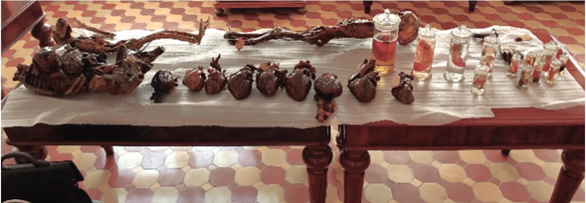
Peter the Great (1672–1725) – the Russian emperor travelled through Europe during the so-called “Great Embassies” to acquire knowledge in the field of science and industrial production. In Amsterdam (Netherlands), he received as a gift from Frederik Ruijsch samples of dried and wet anatomical specimens, consisting of objects of both natural history and human origin, and currently called the “small collection”. The anatomical part of the collection consisted of 11 dry and 13 wet specimens, parts of the human body. The authors rediscovered this collection in 2016 among the exhibits of the fundamental museum of the department of normal anatomy of the Military Medical Academy (St. Petersburg, Russia). In the article we describe the current state of this historical collection, which played an important role in the acquisition of a more extensive collection by Peter the Great from Ruysch for the Kunstkamera (Museum of Anthropology and Ethnography of Peter the Great in St. Petersburg). The Kunstkamera collection is open to the general public, and as a result is well known through publications in literature. On the contrary, the so-called “small collection” was, from the moment of its arrival in Russia in 1698, in the personal possession of Peter the Great. He later divided his small collection for educational purposes between the Aptekerskii Prikaz and the medical school of Nicolaas Bidloo. Around 1800, both parts of the small collection were reunited and transferred to the Medical-Surgical Academy. Today, this collection still serves educational purposes, but is not widely available to the general public. As a result, it remains virtually unknown.
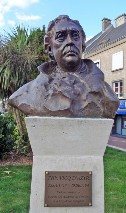
Felix Vicq-d'Azyr (1748-1794) – an encyclopedic scientist of the Enlightenment, a medical practitioner, anatomist, and social reformer, who lived a short, selfless life that ended during the firestorm of the French Revolution. He is one of the founders of comparative anatomy and the discoverer of the phenomenon of homology. Vicq-d'Azyr is known for his contributions to the study of the brain anatomy; some structures of the brain are named after him. Vicq-d'Azyr made an invaluable contribution to the fight against cattle epizootics and to the principles of maintaining sanitary and hygienic standards in high-density residential areas. He was one of the founders of the Royal Society of Medicine of France, as well as the author and co-author of significant medical publications in general therapeutic and surgical areas. His research studies on pathological anatomy, the history of medicine, and ideas for educational reforms are also noteworthy. Although Vicq-d’Azyr lived only 46 years, he left behind an extensive theoretical and practical scientific heritage.


